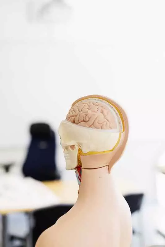
History of anatomical models in the life sciences
Introduction to anatomical models
Anatomical models have played a key role in the life sciences for centuries, providing researchers and students with tools to understand the complexity of living organisms. The use of graphic and physical representations in medical and biological education has been known since ancient times, when scientists sought ways to visualize the structure of the human and animal body.
History of anatomical models in antiquity
The origins of anatomical models date back to times such as Egypt and Greece. In ancient Egypt, doctors were already dissecting cadavers, which allowed them to learn about the structure of the human body. In Greece, thanks to thinkers such as Hippocrates and Galen, more complex methods of depicting anatomy began to be used. Galen, for example, developed a series of drawings to illustrate the workings of various organs.
The Renaissance and the development of anatomical practices
During the Renaissance, interest in the natural sciences increased significantly. Thanks to inventions such as printing, the works of anatomists could be more widely disseminated. Andreas Vesalius, considered the father of modern anatomy, introduced accurate and detailed anatomical illustrations in his work "De humani corporis fabrica," which revolutionized the teaching of anatomy. His work was based on personal research conducted on human bodies, which allowed him to correct many of Galen's erroneous theories.
Revolutions in anatomy of the 17th and 18th centuries
In the 17th and 18th centuries, developments in technology and research methods further developed anatomical models. William Harvey 's discovery of blood circulation marked a turning point in understanding the workings of the circulatory system. The new discoveries also required new models that could clearly represent these complex processes.
This period also saw the beginning of the use of an educational website, where anatomical models were presented to help students better understand body structure. These models were often made of wax or plaster, which allowed for realistic reproduction of anatomical details.
Development of technology in the 19th century
The 19th century was a time of major advances in anatomy and biology. With the development of photographic and microscopic technologies, it became possible to study cellular structures in even greater detail. Rudolf Virchow formulated cell theory, which led to the need for new models depicting cell structure.
During this time, 3D models began to be introduced at medical schools, making it even easier for students to learn anatomy. The use of new materials, such as plastic, made it possible to create more durable and realistic models.
The 20th century and the modern approach to anatomical models
In the 20th century, developments in computer technology brought a revolution in anatomy teaching methods. Computer simulations and specialized software make it possible to teach anatomy interactively, significantly improving student learning. Using technology, students can remotely z(about)ach complete anatomical models, ready for interaction and learning.
Today's anatomical models are not just static sculptures, but also dynamic simulations that present real-time changes, such as blood circulation or the body's response to various stimuli. This new approach greatly facilitates the understanding of complex biological processes.
Summary
The history of anatomical models is a fascinating journey through the centuries, showing how developments in science and technology have affected our understanding of life. From ancient drawings to modern computer simulations, these models have been and continue to be an integral part of the life sciences. Thanks to them, we have been able to understand not only the structure but also the functioning of organisms, which has been crucial to the development of medicine and biology.
The future of anatomical models seems promising. with the development of artificial intelligence and augmented reality, it will be possible to depict the complexity of life even more accurately and intuitively. Who knows what innovations the next decade will bring us in the field of anatomy and anatomical models?